
EMG — Guide to collected data
EMG data was collected on two chimpanzee subjects between March 2012 and June 2015. All EMG recording sessions included 4 channels of EMG recordings, usually from separate muscles but occasionally including multiple recordings of the same muscle. Since the main goal of this project was the documentation of chimpanzee hind limb muscle activity patterns during bipedal walking, the set of four muscles selected for any particular recording session most often was based on proximity. This simplified the process of running the wires along the subject’s limb up to the telemetry transmitter pack that was attached to a harness around the subject’s chest. However, many times an overlying muscle was included when sampling the activity patterns of deeply positioned muscles in order to detect possible electrode tip displacement during the recording session. In addition, the actions of some muscles are related to one another, and therefore some combinations of muscle recordings were selected to be able to observe these interactions.
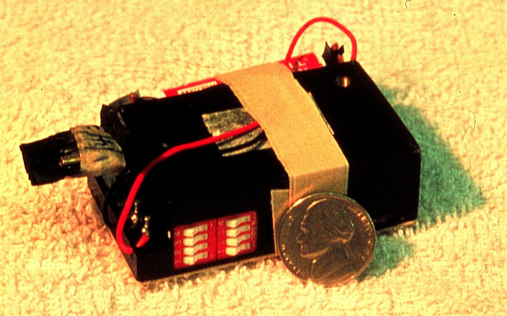
EMG FM telemetry transmitter package (~ 112 g; Bio-Sentry Telemetry). Photograph by Jack Stern, nd
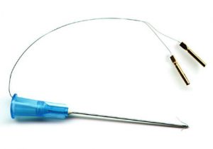
The fine-wire electrode/hypodermic needle assembly used by the Stony Brook Primate Locomotion Lab consisting to two 50 μm nylon insulated wires threaded through a hypodermic needle, prepared in the manner suggest by Basmajian and Stecko (1962). The hypodermic needle is used to position with barred tips of the wires into the muscle belly and then withdrawn, all while the animal research subject is under anesthesia. Photograph by Jack Stern, nd
Prior to positioning the electrodes, the skin over the target muscle was shaved to reduce the chance of the wires catching on the subject’s hair. Following sterilization of the fine-wire electrode/hypodermic needle assemblies in an autoclave, the electrode tips are placed within a muscle belly with a hypodermic needle (which is then withdrawn) while the animal subject is anesthetized with isoflurane gas delivered through a face mask. Following placement of each electrode, back-stimulation is used to determine that it is correctly positioned in the target muscle. After the four electrodes are in place, the wires are guided along the subject’s limb (secured by skin tape) and connected to the telemetry transmitter. Finally, loose clothing was put on the subject to prevent the electrode wires from catching on anything and being pulled out accidently.
Due to our desire to use as minimally invasive methods as possible, we do not attempt to surgically secure the wire electrodes in any fashion. Though this offers the advantage of being able to easily pull the electrodes out at the end of a recording session, it also means that displacement of an electrode is a fairly common occurrence. We therefore monitor the activity of the muscles throughout a recording session to try and detect changes in patterns of recruitment. In addition, we attempt to always complete duplicate recordings of a muscle from each subject to help establish the consistency of a muscle’s activity profile. Like all recordings of biophysical processes, EMG is subject to extraneous noise of unknown origin that usually manifests itself as “movement” or “motion artifact”, that is, low frequency deflections of the baseline sometimes interfering with the higher-frequency EMG signal. Therefore, an individual recording session may be only partially successful.
Catalog of EMG recording sessions
A list of the recordings sessions for the two chimpanzees involved in the Chimpanzee Bipedalism project has been compiled. The recordings are organized by muscle, with recordings for each subject listed separately by date. Each entry includes comments on the quality of the channels, and whether samples of step cycles have been quantified. A variety of behaviors are elicited from the subject by the animal trainer during the recording session, and therefore, samples a quadrupedal walking (G4P) and vertical climbing (VC) as well as bipedalism (G2P) have often been collected. Access to any of these recordings is by permission only.
The EMG data were recorded using ProCapture software package and analyzed using ProAnalyst. An EMG recording session consists of an avi file displaying a continuous view of the subject’s movements during the session, and an au2 file containing the digital EMG data. Both files are opened simultaneously in ProAnalyst to permit correlation of muscle activity patterns to specific motions and behaviors. The avi files can be viewed in other media players, and the au2 files can be opened in a text editor or exel.
If interested in using the EMG data we have collected, please send a description of the nature of your request to Susan.Larson@stonybrook.edu to obtain permission to use this data. With permission you will be given access to password-protected folder containing the list of EMG recording sessions.